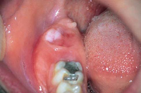
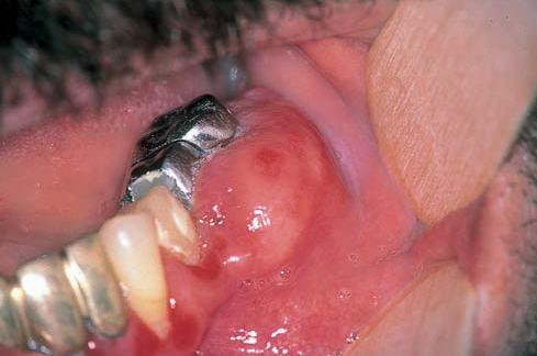
Quantcast
Channel: Dentistry lectures for MFDS/MJDF/NBDE/ORE

A NOTE ON DENTAL CALCULUS

$
0
0

Dental calculus


For the periodontal diseases:

  •     The primary etiologic factor is: Is the dental plaque.
  •       The associated factor: is the dental calculus, it helps in new formation of the plaque.
  •       The modifying factor: is a systemic disease, it aggravates the disease when the plaque is presents.


Calculus:
  •    Is a mineralized dental plaque that occurs in the tooth surfaces & dental prosthesis, it has many forms:
  •    Bridging over the gingival margin.
  •     Follow the festooning shape of the dentition.
  •     Lobular form.
  •     In case of malalignment :àprotected area for the plaque à calculus

 Classification:
             Supragingival                  &               subgingival calculus..

         Generally: both can occur together or one may appear alone.


Subgingival calculus: 
  •      Gingival fluid origin.
  •       Below the crest of the gingival margin.
  •       Hard, dark& flint like.
  •       Greenish black or dark brown in color.
  •       Firmly attached to the tooth, can’t be seen and detected by explorer No.621 probe.
  •       Extent nearly to the base of the pocket in chronic periodntitis, but doesn’t reach the Junctional epithelium.

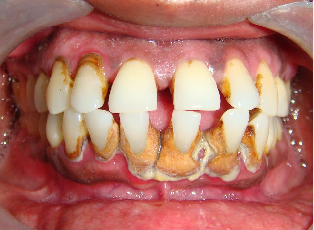
 Supragingival calculus:    
  •     Saliva origin.
  •    Coronal to the gingival margin. Can be composed of supra &sub gingival calculus.
  •    Hard, clay like consistency, White, white yellowish in color& its color may be affected by the tobacco or food stain.
  •     Easy to be seen in the oral cavity, may be generalized or localized.
  •     Easy to be removed &usually recurrent especially in the: Lower incisors. 
  • Most common location :near to the orifices of the S. glands’ ducts

Parotid gland’s duct ”stenson”----->opposite to buccal surface of maxillary molars.


Submandibular “bartholine” & sublingual “wharton” ducts ----->Lingual surface of lower incisors.

it’s shape : either covers the occlusal surfaces or  bridge like structure over interdental papilla.


                                                Calculus contents:


Inorganic contents:70-90%


        Organic contents


2/3 of the calculus inorganic component is in crystalline form ;there are  4 types of crystals .


The crystals are:

hydroxyappatite ,58% à

magnesium white locate,21%  àmost in post

octacalcium phosphate,12% Brushite, 9% àmost in mandibular anteriors.

Detected more frequently in supragingival calculus.

Constitute the bulk.


Generally 2 or more crystals are detected in the calculus.


Incidence of 4 crystals à varies with age of calculus.


Mixture of  :

Protein-poly saccharide complex + desquamated host cells (leukocytes & host cells) + microorganisms.


Carbohydrates (1.9-9.1%):

Glucose , glactose   ,mannose ,arabinose ,rhamnose glucoric acid ,glactouric acid glucoseamine & glactose amine. à all are present in saliva except :

Rhaminose  &  arabinose .


Salivary proteins (5.9-8.2%):

Most are amino acids.


lipids 0.2%:nutral fat ,fatty acids ,cholesterol ester, phospholipids & cholesterol.


*Contents:

          The differences

    Supra gingival calculus

Between:

Sub gingival calculus


hydroxyappatite:

Ca Ph :

Mg white:

brushite:

ratio of Ca/Ph:

sodium contents:


salivary proteins:

             Equal.  

More.

Less.

More.

Low.  

Increase with the depth of PD pocket.

Yes                

            Equal

Less.

More

Less.

Higher.


No.


Calculocementum:

Is the calculus has morphological appearance similar to cementum. This is because the calculus is interdigitates the cementum & no differences between them.


Mode of attachment of the calculus to the tooth surface:

  • Close adaptation under surface depression.
  • In sub gingival calculus.
  • By organic pellicle (very weak)
  • Penetration of the bacteria to the cementum.
  • Mechanical interlocking to the surface irregularities: resorption lacuna or caries, in the cementum by sharpies fibers.                          

Calculus formation:

Calculus is the dental plaque that undergoes mineralization.

Calcification starts                     4-8 hrs   after plaque.

50 % become mineralized after   2 days.

60-90                                         12 days.
  • Plaque can be daily removed at home by brushing but the calculus is
  • not ,it is only removed clinically by the dentist .
  • Calculus formation à the bacterial action will stopped (adv) but it will act as stagnation area for new plaque accumulationà (protection for plaque). 
  • Early plaque contains small amount of inorganic material but it will increase as the plaque develops into calculus. 
  • All plaque doesn’t necessarily undergo calcification.
  • It reaches a plateau of maximal mineral by 2 days.
  • Microorganisms are not always essential in calculus formation. 
  • Plaque has ability to conc. The Ca at  2-20 times it’s level in saliva. 
  • There is a suggestion that Ph is more critical than Ca in plaque mineralization.
  • Early plaque of heavy former àmore Ca ,3 times Ph &less K than non calculus former.


A NOTE ON CLASSIFICATION, CLINICAL FEATURES, PATHOGENESIS OF PERIODONTAL POCKET

$
0
0

The periodontal pocket, defined as a pathologically deepened gingival sulcus, is one of the most important clinical features of periodontal disease. All different types of periodontitis share histopathologic features such as tissue changes in the periodontal pocket, mechanisms of tissue destruction, and healing mechanisms. They differ, however, in their etiology, natural history, progression, and response to therapy.


CLASSIFICATION

Deepening of the gingival sulcus may occur by coronal movement of the gingival margin, apical displacement of the gingival attachment, or a combination of the two processes.

Illustration of pocket formation indicating expansion in two directions (arrows) from the normal gingival sulcus (left) to the periodontal pocket (right)
Different types of periodontal pockets. A, Gingival pocket. There is no destruction of the supporting periodontal tissues. B, Suprabony pocket. The base of the pocket is coronal to the level of the underlying bone. Bone loss is horizontal. C, Intrabony pocket. The base of the pocket is apical to the level of the adjacent bone. Bone loss is vertical.
Pockets can be classified as follows:

Gingival pocket (pseudo pocket):

This type of pocket is formed by gingival enlargement without destruction of the underlying periodontal tissues. The sulcus is deepened because of the increased bulk of the gingiva


Periodontal pocket:

This type of pocket occurs with destruction of the supporting periodontal tissues. Progressive pocket deepening leads to destruction of the supporting periodontal tissues and loosening and exfoliation of the teeth.

Two types of periodontal pockets exist:

Suprabony(supracrestal or supraalveolar), in which the bottom of the pocket is coronal to the underlying alveolar bone.


Intrabony(infrabony, subcrestal or intraalveolar), in which the bottom of the pocket is apical to the level of the adjacent alveolar bone. In this second type, the lateral pocket wall lies between the tooth surface and the alveolar bone.


Pockets can involve one, two, or more tooth surfaces and can be of different depths and types on different surfaces of the same tooth and on approximating surfaces of the same interdental space . Pockets can also be spiral (i.e., originating on one tooth surface and twisting around the tooth to involve one or more additional surfaces). These types of pockets are most common in furcation areas.


Classification of pockets according to involved tooth surfaces. A, Simple pocket. B, Compound pocket. C, Complex pocket
CLINICAL FEATURES

Clinical signs such as bluish-red, thickened marginal gingiva;

A bluish-red vertical zone from the gingival margin to the alveolar mucosa;

Gingival bleeding, suppuration,or both;

Tooth mobility;

And diastema formation and symptoms such as localized pain or pain "deep in the bone" are suggestive of the presence of periodontal pockets.

The only reliable method of locating periodontal pockets and determining their extent is careful probing of the gingival margin along each tooth surface.

A, Extrusion of the central incisor and diastema associated with the periodontal pocket. B, The entire length of the periodontal probe inserted to the base of the periodontal pocket in the central incisor
Correlation of Clinical and Histopathologic Features of the Periodontal Pocket

Clinical Features

Histopathologic Features

1. The gingival wall of the periodontal pocket presents various degrees of bluish-red discoloration; flaccidity; a smooth, shiny surface; and pitting on pressure.

1. The discoloration is caused by circulatory stagnation; the flaccidity, by destruction of the gingival fibers and surrounding tissues; the smooth, shiny surface, by the atrophy of the epithelium and edema; the pitting on pressure, by edema and degeneration.

2. Less frequently, the gingival wall may be pink and firm.

2. In such cases, fibrotic changes predominate over exudation and degeneration, particularly in relation to the outer surface of the pocket wall. However, despite the external appearance of health, the inner wall of the pocket invariably presents some degeneration and is often ulcerated.
3. Bleeding is elicited by gently probing the soft tissue wall of the pocket.

3. Ease of bleeding results from increased vascularity, thinning and degeneration of the epithelium, and the proximity of the engorged vessels to the inner surface.

4. When explored with a probe, the inner aspect of the periodontal pocket is generally painful.

4. Pain on tactile stimulation is due to ulceration of the inner aspect of the pocket wall.


5. In many cases, pus may be expressed by applying digital pressure.


5. Pus occurs in pockets with suppurative inflammation of the inner wall.


On the basis of depth alone, however, it is sometimes difficult to differentiate between a deep normal sulcus and a shallow periodontal pocket. In such borderline cases, pathologic changes in the gingival distinguish the two conditions.


PATHOGENESIS

The initial lesion in the development ofperiodontitis is the inflammation of the gingiva in response to a bacterial challenge. Changes involved in the transition from the normal gingival sulcus to the pathologic periodontal pocket are associated with different proportions of bacterial cells in dental plaque. Healthy gingiva is associated with few microorganisms, mostly coccoid cells and straight rods. Diseased gingiva is associated with increased numbers of spirochetes and motile rods. However, the microbiota of diseased sites cannot be used as a predictor of future attachment or bone loss because their presence alone is not sufficient for disease to start or progress.

Extension of the junctional epithelium along the root requires the presence of healthy epithelial cells. Marked degeneration or necrosis of the junctional epithelium retards rather than accelerates pocket formation. Degenerative changes seen in the junctional epithelium at the base of periodontal pockets are usually less severe than those in the epithelium of the lateral pocket wall. Because migration of the junctional epithelium requires healthy, viable cells, it is reasonable to assume that the degenerative changes seen in this area occur after the junctional epithelium reaches its position on the cementum.

The transformation of a gingival sulcus into a periodontal pocket creates an area where plaque removal becomes impossible, and the following feedback mechanism is established:


The rationale for pocket reduction is based on the need to eliminate areas of plaque accumulation.


A Note on Bone Swellings in Jaw Bones

$
0
0

Bone Swellings

Bone swellings are lesions that characteristically present as asymptomatic hard lumps, covered by normal epithelium. Developmental disorders, benign and malignant tumors are included in this group of lesions.

  •        Torus mandibularis
  •        Torus palatinus
  •        Multiple exostoses
  •        Osteoma
  •        Osteosarcoma
  •         Chondrosarcoma
  •         Burkitt lymphoma
  •         Multiple myeloma
  •         Paget disease
  •       Odontogenic tumors

Torus Mandibularis

Definition and etiology Torus mandibularis is a developmental malformation of unknown etiology.

Clinical features It presents as an asymptomatic bony swelling, covered by normal mucosa. The lesion displays slow growth during the second and third decades of life. Characteristically, the lesions appear bilaterally on the lingual surface of the mandible, usually in the area adjacent to the bicuspids. The diagnosis is based on clinical criteria.

Torus mandibularis
Treatment Unnecessary unless full denture construction is required.


Torus Palatinus

Torus palatinus at the midline of the hard palate
Definition and etiology Torus palatinus is a developmental malformation of unknown etiology.

Clinical features It presents as a slow-growing, nodular, lobular or spindled, asymptomatic bony swelling covered by normal mucosa. Characteristically, the lesion appears along the midline of the hard palate.It occurs more often in women, and usually appears during the third decade of life. The diagnosis is based on the clinical findings.

Treatment Unnecessary unless full denture construction is required.


Multiple Exostoses

Multiple exostoses may occur on the buccal surface of the maxilla, and rarely on the mandible. Clinically, the lesions appear as multiple asymptomatic bony swellings. The diagnosis is based on the clinical findings.

Multiple exostoses on the maxilla.
Treatment Unnecessary unless full denture preparation is required.


Osteoma

Definition Osteoma is a benign neoplasm that consists of mature compact or cancellous bone.

Etiology Unknown.

Clinical features

It presents as an asymptomatic, slow-growing bony swelling of the jaws. The size ranges from a few millimeters to several centimeters. Multiple jaw osteomas are a common feature of Gardner syndrome.

Gardner syndrome: osteoma of the mandible.
Laboratory tests Histopathological examination, radiography.

Differential diagnosis Exostoses, osteosarcoma.

Treatment Surgical excision.


Osteosarcoma

Definition Osteosarcoma is the most common primary malignant neoplasm of bone.

Etiology Unknown.

Clinical features

The jaws are affected in 6–7% of cases, and usually during the third decade of life. Both jaws are affected equally and it is more common in men. Clinically, the lesion presents as a rapidly growing hard swelling that progressively produces facial deformity. Pain, paresthesia, tooth loosening, and nasal obstruction may also occur.

Osteosarcoma of the upper jaw, presenting as a hard swelling.
Laboratory tests Histopathological examination, radiography, CT scans.

Differential diagnosis Chondrosarcoma, Ewing sarcoma, metastatic tumors, odontogenic tumors and cysts, giant-cell tumor.

Treatment Surgical excision and supplementary radiotherapy and chemotherapy.


Chondrosarcoma

Chondrosarcoma  is more common in men than in women, between 30 and 60 years of age. Clinically, it appears as a painless hard swelling that progressively enlarges, causing extensive bone destruction with pain and loosening of the teeth.


Burkitt Lymphoma

Definition Burkitt lymphoma is a high-grade malignant B-lymphocyte lymphoma.

Etiology Epstein–Barr virus is closely associated.

Clinical features

The malignancy is prevalent in central Africa (the endemic form), and usually affects children 2–12 years of age. Cases have also been observed in other countries (the nonendemic form), and recently in patients with AIDS. The jaws are the most common site of lymphoma (60–70%). Clinically, it presents as a rapidly growing hard swelling that causes bone destruction, tooth loss, and facial deformity.Pain, paresthesia and large ulcerating or nonulcerating masses may also be seen.

Burkitt lymphoma, facial deformity.
Burkitt lymphoma, gingival mass
Burkitt lymphoma on the gingiva in a young patient with AIDS
Laboratory tests Histopathological examination, radiography.

Differential diagnosis Central giant-cell granuloma, ossifying fibroma, other non-Hodgkin lymphomas, and odontogenic tumors.

Treatment Chemotherapy, radiotherapy.


Multiple Myeloma

Definition Multiplemyeloma is a relatively rare malignant plasma-cell disorder.

Etiology Unknown.

Clinical features The malignancy is more common in men over 50 years of age, and the jaws are affected in about 30% of cases. Clinically, it presents with bone swelling, tooth mobility, pain, and paresthesia. A painless soft swelling, usually on the alveolar mucosa and gingiva, may develop as part of the overall disease spectrum.

Multiple myeloma, swelling on the gingiva
Laboratory tests Bone-marrow biopsy, radiography, serum and urine protein electrophoresis.

Differential diagnosis Plasmacytoma, non-Hodgkin lymphoma, Ewing sarcoma, leukemia, Langerhans cell histiocytosis.

Treatment Chemotherapy, radiotherapy.


Paget Disease

Definition Paget disease, or osteitis deformans, is a chronic, relatively common disorder characterized by uncoordinated bone resorption and deposition.

Etiology Unknown.

Clinical features Clinically, the signs and symptoms develop gradually and are characterized by bone pain, headache, deafness, visual disorders, dizziness, and progressive bone enlargement. Progressive expansion of the maxilla and the mandible lead to symmetrical thickening of the alveolar ridges.

Paget disease, enlarged maxilla
Edentulous patients may complain that their dentures do not fit due to alveolar enlargement.

Paget disease, alveolar enlargement
Delayed wound healing, bleeding, and osteomyelitis after tooth extraction may occur. The maxilla is more frequently affected than the mandible. Malesare more often affected than females. Two major forms of the disease are recognized: (a) the monostotic, and (b) the polyostotic. The clinical diagnosis should be confirmed by a histopathological and radiographic examination. Elevations of serum alkaline phosphatase and urinary hydroxyproline levels are common findings.

Differential diagnosis Fibrous dysplasia, osteosarcoma, multiple exostoses, fibro-osseous lesions.

Treatment Most cases require no treatment. Calcitonin and bisphosphonates may slow the pathological process.

Odontogenic Tumors
Definition Odontogenic tumors are a group of lesions that originate from odontogenic epithelium and ectomesenchyme.

Etiology Unknown. Some are neoplasms and others hamartomas.

Classification On the basis of the tissue of origin, three major varieties are recognized: (a) tumors of odontogenic epithelium, (b) tumors of odontogenicectomesenchyme, and (c) mixedod ontogenic tumors.

Clinical features Most odontogenic tumors are usually asymptomatic for long time and are discovered only during a routine radiographic examination. However, with time they may form a usually painless slow-growing swelling or expansion of the mandible or the maxilla.

Odontogenic myxoma, expansion of the retromolar area
Extraosseous calcifying epithelial odontogenic tumor presenting as a gingival mass
The clinical signs and symptoms are not diagnostic and the final diagnosis should be made by radiographic and histopathological examinations.

Differential diagnosis Different varieties of odontogenic tumors, odontogenic cysts, osteosarcomas, chondrosarcomas, multiplemyeloma.

Treatment Surgical excision.

A NOTE ON SPACE MAINTENANCE IN THE PRIMARY DENTITION

$
0
0

SPACE MAINTENANCE IN THE PRIMARY DENTITION


Space maintenance can be defined as the provision of an appliance (active or passive) which is concerned only with the control of space loss without taking into consideration measures to supervise the development of dentition.

Space maintainers are appliances used to maintain space or regain minor amounts of space lost, so as to guide the unerupted tooth into a proper position in the arch.

IDEAL REQUIREMENTS

  • It should maintain the entire mesio-distal space created by a lost tooth.
  • It must restore the function as far as possible & prevent over-eruption of opposing teeth.
  • It should be simple in construction.
  • It should be strong enough to withstand the functional forces.
  • It should not exert excessive stress on adjoining teeth.
  • It must permit maintenance of oral hygiene.
  • It must not restrict normal growth & development and natural adjustments which take place during the transition from deciduous to permanent dentition.
  • It should not come in the way of other functions.


A NOTE ON PTERYGOPALATINE FOSSA

$
0
0

PTERYGOPALATINE FOSSA


The pterygopalatine fossa—

           A small, pyramid-shaped space.

           Situated between the maxilla, sphenoid, and palatine bones.

           It communicates via canals, fissures, and foramina with various regions of the skull.

          The contents of the pterygopalatine fossa include

                     The terminal portion of the maxillary artery;

                     The pterygopalatine ganglion;

                     The maxillary division of the trigeminal nerve; and branches of these structures.

Maxillary Artery

The third, or pterygopalatine portion, of the maxillary artery enters the pterygopalatine fossa from the infratemporal fossa via the pterygomaxillary fissure

Maxillary artery and its distribution in the deep face

Branches of the pterygopalatine portion of the maxillary artery are the posterosuperior alveolar, infraorbital, greater palatine, pharyngeal, and sphenopalatine arteries as well as the artery of the pterygoid canal.

The posterior superior alveolar arterybranches from the maxillary artery as that vessel enters the pterygomaxillary fissure. It travels on the maxillary tuberosity and enters the posterior superior alveolar foramen accompanied by the like-named nerve. The vessel ramifies within the maxilla to vascularize the maxillary sinus, molars, and premolars as well as the neighboring gingiva.

The infraorbital artery, a continuation of the maxillary artery, enters the orbit through the inferior orbital fissure, lies in the infraorbital groove, leaves the orbit via the infraorbital canal, and enters the face by way of the infraorbital foramen. Branches of the infraorbital artery are the orbital branches, serving the lacrimal gland and the inferior oblique and inferior rectus muscles; the anterior superior alveolar branches, which vascularize the anterior teeth and the maxillary sinus; and the facial branches.

The greater palatine artery and its branch, the lesser palatine artery, pass through the pterygopalatine canal and gain entrance to the palate via the greater palatine and lesser palatine foramina, respectively, to vascularize the hard and soft palates as well as associated structures. The pharyngeal branch passes dorsally, through the pharyngeal canal, to vascularize the auditory tube, sphenoidal sinus, and portions of the pharynx. The sphenopalatine artery leaves the pterygopalatine fossa via the sphenopalatine foramen on its medial wall to enter the nasal fossa. The distribution of this vessel and its branches is discussed later in this chapter. The small artery of the pterygoid canal passes through the posterior wall of the pterygopalatine fossa via the pterygoid canal. It supplies part of the auditory tube, pharynx, middle ear, and sphenoidal sinus.

Maxillary Nerve


The maxillary division of the trigeminal nerve enters the pterygopalatine fossa at its posterior boundary via the foramen rotundum. While in the fossa, it gives off the zygomatic nerve, which, passing into the orbit through the inferior orbital fissure, will bifurcate to form the zygomaticotemporal and zygomaticofacial nerves.

The maxillary division of the trigeminal nerve
The posterior superior alveolar nerves also branch from the maxillary nerve, exit the fossa via the pterygomaxillary fissure, and enter the maxillary tuberosity to serve the maxillary sinus, molars, and adjacent gingiva and cheek. The maxillary nerve then enters the orbit by way of the inferior orbital fissure and is referred to as the infraorbital nerve.

While in the pterygopalatine fossa, the maxillary nerve communicates with the pterygopalatine ganglion via two small trunks, the pterygopalatine nerves; however, these nerves do not bear a functional relationship with the ganglion. Postganglionic parasympathetic fibers derived from the ganglion ride along and distribute with branches of the maxillary division of the trigeminal nerve.

Pterygopalatine ganglion and associated nerves and arteries
Orbital branches are slender nerves that supply the periosteum of the orbit and the mucoperiosteum of the ethmoidal and sphenoidal sinuses. The greater palatine nerve and its branches, the lesser palatine and posterior inferior nasal branches, descend through the pterygopalatine canal to supply regions of the palate, gingiva, tonsil, and lateral wall of the nasal fossa.

Posterior superior nasal branches leave the pterygopalatine fossa via the sphenopalatine foramen to serve the posterior aspect of the nasal fossa and part of the ethmoidal sinus. Its nasopalatine branch grooves the vomer bone in its path to the incisive foramen of the anterior hard palate, which it supplies. The pharyngeal nerve traverses the pharyngeal canal to innervate part of the nasopharynx.


Pterygopalatine Ganglion

The pterygopalatine ganglion seems to be functionally associated with the maxillary division of the trigeminal nerve because it is suspended by the pterygopalatine nerves within the fossa. It is, however, a parasympathetic ganglion of the facial nerve (cranial nerve VII).

This ganglion receives its parasympathetic preganglionic root by way of the pterygoid canal, which opens onto the posterior wall of the fossa. The preganglionic parasympathetic fibers synapse with postganglionic parasympathetic cell bodies within the ganglion. Postsynaptic parasympathetic fibers leave the ganglion and distribute with branches of the maxillary division of cranial nerve V. These fibers are secretomotor in function. They provide parasympathetic flow to the lacrimal gland and mucosal glands of the nasal fossa, palate, and pharynx.


Methods for Removal of root filling material in preparation for posts

$
0
0

Methods for Removal of root filling material in preparation for posts


Obturation techniques

Gutta-percha is today the universally accepted core material used for root canal obturation. However, the techniques for placement differ, but all require the use of sealing cement. Obturation techniques include cold lateral compaction of gutta-percha points, compaction of gutta-percha that has been heat softened in the canal and compacted (eg System B), thermoplastisised gutta-percha which is injected into the canal (eg Obtura and UltraFil) and finally compaction of gutta-percha which has been placed in the canal and softened by mechanical means (eg McSpadden compactors). These obturation techniques are unlikely to have an impact on the final apical seal once post space preparation has been carried out. An alternative obturation technique also exists which involves heated gutta-percha surrounding a plastic or metal carrier (eg Thermafil). The carrier ensures that the gutta-percha passes to the correct working length and is left in situ with the gutta-percha. During mechanical gutta-percha removal of this system, there is greater potential to disrupt the apical gutta-percha and a number of dye leakage studies have supported this hypothesis. It would therefore be prudent not to use such obturation techniques if placement of a post is anticipated.


Chemical removal

Solvents such as oil of eucalyptus, oil of turpentine and chloroform have been used to soften gutta-percha for removal, with the latter two being the most effcicient. However, some of these materials and especially chloroform are hazardous to use as they are toxic and potentially carcinogenic. Oil of turpentine is less toxic, but there is concern that solvents in general lead to a dimensional change in the gutta-percha, leading to increased microleakage. This together with the fact that it is difficult to control the depth of softening of the gutta-percha and potential leakage of the solvents into the periradicular tissues should be sufficient to discourage their use for gutta-percha removal for post placement. They are however a necessary adjunct in root canal re-treatment cases.


Thermal removal

A heated instrument such as a lateral compactor can be inserted into the gutta-percha to the desired length to soften and remove the gutta-percha. However, in narrow canals, fine instruments lose their heat quickly and gutta-percha removal can be difficult. A System B spreader is ideal for removal of gutta-percha.

System B with heated plugger (200°C) in foreground, with rubber stop placed at the desired length for gutta-percha removal  
From a pre-operative radiograph a plugger should be chosen of the correct dimensions that is likely to bind at the desired post length and this position should be marked on the plugger with a rubber stop. The tip should be placed in the gutta-percha and with the heat applied driven slowly to the desired post length in about 2–3 seconds. The heat should be removed and the plugger allowed cooling for about 7–10 seconds, twisted and then removed with the coronal gutta-percha. Alternatively, a short burst of heat to the plugger will allow for easy removal. It is important that the plugger is sufficiently hot to completely soften the gutta-percha. If too cool, it will result in the gutta-percha remaining sticky with the risk of dislodging the apical gutta-percha. An instrument such as a Buchanan plugger can then be used to vertically compact the softened gutta-percha. Such a technique is useful in removing old gutta-percha which can become quite hard.

Buchanan plugger  
Some authors would suggest that gutta-percha should be removed with heated techniques as a routine and mechanical removal only used if heat is insufficient.If mechanical removal is used, a heated instrument can be used to soften the most coronal gutta-percha, so that it can be vertically compacted and adapted to the canal walls to create a seal.


Mechanical removal

Mechanical removal of gutta-percha is efficient and probably the most commonly used technique, but it is a technique that can result in the most damage to tooth tissue. If done incorrectly, it can weaken the root unnecessarily, damage the periodontium and in some cases lead to root perforation.

Periapical radiograph showing teeth 13 and 12 used as double abutments for a fixed-fixed conventional bridge 

 Periapical radiograph of tooth 23 with a post that poorly fits the prepared post hole 
A non-end cutting bur such as a Gates-Glidden or Peeso reamer should be used for gutta-percha removal, as these will cut and remove the relatively softer gutta-percha preferentially to the dentine of the canal walls.


The sequence in which the burs are used is important so that a rise in temperature at the root surface, which could damage periodontal cells, is avoided and the risk of preferentially cutting away root dentine to one side of the root canal is reduced.

 Periapical radiograph of root filled central incisor teeth (top left) from which the diameter of the post to be used can be estimated  
Temperature rise on the root surface has been investigated in a number of studies. A Gates-Glidden bur rotating at 8,000 rev min-1 results in a small rise in temperature at the root surface. However, both tapered and parallel-sided post drills produce a significant increase in temperature in excess of 17°C. Peeso reamers also generate significant rises in temperature, higher than that reached with Gates-Glidden burs and Parapost twist drills. To reduce this temperature increase, which could potentially damage cells in the periodontal ligament, it is important that the smaller sized Gates-Glidden burs are used first, working up through the sizes in turn, until no gutta-percha is removed apically. At this stage the smallest post drill can be used, again working up through the size sequence until the final post size is reached.

A note on Jacket crowns, Indications, Alternatives, Assessment of teeth and Tooth preparation

$
0
0

A note on Jacket crowns
Indications

Discolored teeth


Fractured teeth


Grossly carious teeth


Hypoplastic teeth


Tooth wear


As part of a bridge


Alternatives to JCs

Bleaching


Composite veneering


Porcelain veneering


Composite restorations

Resin bonded bridges

Assessment of Individual teeth

Vitality / status of the pulp

Size of the pulp in vital teeth

Resting lip line and smile line

Aesthetics

Pre operative work up

Finalize the material/s

Depth of preparation

Path of insertion

Appropriate burs, trays and materials

Local anesthesia if tooth is vital

Shade selection


Stages of tooth preparation

Depth orientation grooves

           Labial, incisal reduction


              Interdental reduction


           Palatal cervical collar


            Cingulum reduction


              Gingival margin


A Note on Anterior cross bite

$
0
0

Anterior cross bite

Possible causes

Class III skeletal pattern


Crowding


Retained primary teeth and roots


Presence of supernumerary teeth


Trauma


Clinical features

  • Instanding maxillary incisor occluding behind the corresponding lower incisors.
  • Over bite which can vary from nothing to excessive depth.
  • Gingival recession of the lower incisor involved.
  • Forward displacement of the  mandible-  instanding tooth comes into premature contact along the normal path of closure.
  •      mobility of the lower incisor involved in the cross bite.

Methods available for correction

Spoon handle biting.

Removable appliance with either cantilever spring or screw.

Lower inclined bite plane.

     
Spoon handle biting

Instruct the child to bite on a spoon handle to guide the incisor which is erupting into cross bite.

 Do not attempt this on an erupted incisor which has a positive over bite.

Once cross bite established,

Identify predisposing factors and remove them to prevent development of the cross bite

Once the cross bite is established, identify and remove aetiological factors responsible and select appropriate method for correction.


Removable appliance

Selection of appliance and the spring design depend on,

      Axial inclination of the tooth


Depth of the over bite.


Amount of forward movement required


Number of teeth involved in the cross bite


Appliance design

Select the best active component which should be used depending on the above factors discussed.

Adequate number of clasps

Add adequate number of clasps to the appliance to resist the reaction of the vertical component of the active force.

Appliance design

Add posterior bite plane to disocclude teeth

Instruction to the technician

Adams clasps on 6/6 d /d

Double cantilever spring(Z spring) on /1

Posterior bite plane (half molar capping)


Management of patient

Fit the appliance and give adequate instructions to the patient to wear it regularly including meal time.

Activate the spring by the correct amount.

Adjust clasps and check the thickness of the molar capping.

Give adequate instruction to the parents as well.

Monitor progress of treatment

If cross bite is corrected and depth of the over bite adequate to ensure the stability of the occlusion discontinue appliance

Monitor the development of the occlusion until the occlusion of the permanent dentition is established.

Incisors cross bite before and after correction


Incisor cross bite not corrected early lead to a severe malocclusion 


A NOTE ON ANATOMY OF PAROTID GLAND

$
0
0

Parotid gland


The parotid gland is the largest of the salivary glands.


The parotid, a serous compound tubulo-alveolar gland, is yellowish, lobulated, and irregular in shape.


It occupies the interval between the sternomastoid muscle and the mandible.


Average Wt - 25gm (varies in weight from 14 to 28 gm)


Surface anatomy

The parotid gland lies inferior to the zygomatic arch, anteroinferior to the external acoustic meatus, anterior to the mastoid process, and posterior to the ramus of the mandible.


Relations


The parotid gland is enclosed in a sheath (parotid fascia) and is shaped roughly like an inverted pyramid, with three (or four) sides (fig A).

It has a base (from which the superficial temporal vessels and auriculotemporal nerve emerge),apex (which descends inferior and posterior to the angle of the mandible),and lateral, anterior, and posterior (or posterior and medial) surfaces.

The lateral surface is superficial and contains lymph nodes.

The anterior surface is grooved by the ramus of the mandible and masseter (fig.B), producing a medial lip (from which the maxillary artery emerges) and a lateral lip, under cover of which the parotid duct, branches of the facial nerve, and the transverse facial artery emerge (see fig. C).


The posterior surface is grooved by  the mastoid process and the sternomastoid and digastric muscles and  more medially by the styloid process and its attached muscles.


Medially, the superior part of the gland is pierced by the facial nerve and the inferior part by the external carotid artery.


The following structures lie partly within the parotid gland, from superficial to deep:


1. The facial nerve forms the parotid plexus within the gland and separates the glandular tissue partially into superficial and deep layers ("lobes"). In surgical excision of the parotid gland (e.g., for a tumor), damage to the facial nerve is a possibility.


2. The superficial temporal and maxillary veins unite in the gland to form the retromandibular vein, which contributes in a variable manner to the formation of the external jugular vein (see fig. D).


3. The external carotid artery divides within the parotid gland into the superficial temporal and maxillary arteries.


Parotid duct

The parotid duct is about 7 cm long


The parotid duct, emerging under cover of the lateral surface, runs anteriorward on the masseter and turns medially to pierce the buccinator.


The branching of the duct can be examined radiographically after injection of a radio-opaque medium. 


The parotid duct, which is palpable, opens into the oral cavity on the parotid papilla opposite the upper second molar tooth.


Innervation of parotid gland( fig. E)


Preganglionic parasympathetic secretomotor fibers (from the glossopharyngeal, tympanic, and lesser petrosal nerves) synapse in the otic ganglion.


Postganglionic fibers travel with the auriculotemporal nerve and so reach the gland.


Cranial nerves VII and IX communicate, so that secretory fibers to each of the three major salivary glands may travel in both the facial and glossopharyngeal nerves.


The sympathetic supply to the salivary glands includes vasomotor fibers.


Blood supply


The arteries supplying the parotid gland are derived from the external carotid, and from the branches given off by that vessel in or near its substance. The veins empty themselves into the external jugular, through some of its tributaries.


Lymphatics


The lymphatics end in the superficial and deep cervical lymph glands, passing in their course through two or three glands, placed on the surface and in the substance of the parotid.


Best of five Review - Angular cheilitis

$
0
0

Angular cheilitis


Angular cheilitis is a combined staphylococcal, streptococcal, and candidal infection, involving the tissues at the angle of the mouth, often with an underlying precipitating factor, e.g. iron deficiency and B12 deficiency anaemia. Therefore, haematological deficiency should be investigated with a FBC red cell folate, B12, and glucose.


Anecdote suggests aninadequate OVD can also predispose, but correction of this alone will not resolve the condition. Often associated with chronic atrophic candidosis. Clinically, see red, cracked, macerated skin at angles of the mouth, often with a gold crust.


Infecting organisms can be identified on culture ofswabs of the area, although it is usual to make a clinical diagnosis.


Rx:miconazole cream, which is active against all three infecting organisms. Rx needs to be prolonged, up to 10 days after resolution of clinical lesion, and carried out in conjunction with elimination of any underlying factors.


Unless the classic golden yellow crusts associated with S. aureus are present, treatment should be commenced withantifungal drugs, e.g. a combined miconazole/hydrocortisone cream (miconazole has some antibacterial properties).


When clinical features indicateS. aureus infection, fusidic acid cream is appropriate. If intra-oral candidiasis is present, this must be treated concurrently or recurrence of the angular stomatitis will occur. Iron deficiency is a significant aetiological factor in angular cheilitis.


Patterson–Brown-Kelly syndrome (Plummer–Vinson syndrome)

Dysphagia (due to a post-cricoid candida web), microcytic

hypochromic anaemia, koilonychia and angular cheilitis (secondary to the anaemia)

best of five in dentistry review

$
0
0

Recurrent herpes(herpes labialis)

                                                  Giant cell arteritis


                                              Burning mouth syndrome


Trigeminal Neuralgia mcq review

Bilateral parotid swelling MCQ review

Orofacial granulomatosis MCQ review

giant cell arteritis


Kaposi's sarcoma

ANTIFUNGAL AGENTS

$
0
0

ANTIFUNGAL  AGENTS

Most fungal infections in the oral cavity are due to Candida species,
most commonly Candida albicans.
Where candidosis is related to dentures, denture hygiene  instruction should be stressed. Non-metal dentures should be soaked regularly overnight in sodium hypochlorite 1% (Milton’s solution) and metal-containing dentures similarly in chlorhexidine 0.2% solution.
Nystatin and amphotericin (polyenes)
These agents attach to the fungal cell membrane and disrupt fluid and electrolyte permeability. They are not absorbed from the GI tract and hence act locally .
        Dosage regimens for nystatin and amphotericin
Nystatin
Pastilles 100 000 units
Oral suspension 100 000 units/ml
Ointment/cream 100 000 units/g
Amphotericin
Lozenges 10mg
Oral suspension 100 mg/ml
Miconazole (an imidazole)
Similar action to the polyenes. Effective against some Gram-positive
bacteria such as Staph. aureus. More effective than polyenes in angular cheilitis due to possible mixed fungal/bacterial infection.
Available as oral gel, cream and in combination with hydrocortisone.
Oral gel (25 mg/ml) 5–10 ml held over area affected (after food) or applied to fitting surface of upper denture for the treatment of denture stomatitis (chronic erythematous candidosis).
Cream (2%) Apply to angles of lips 2–3 times daily. Cream or ointment (2%) with hydrocortisone (1%) Apply to angles of lips 2–3 times daily. May be useful for clearing long-standing angular cheilitis but should not be used for longer than 10 days.
Fluconazole (a triazole)
This systemically acting agent inhibits fungal enzymes concerned
with ergosterol synthesis. It appears to have low systemic toxicity.
Form Capsules (50 mg) and oral suspension (50 mg/5 ml). Dose 50 mg daily for 7–14 days. Higher doses will be required in immune compromised patients.
Cautions Avoid in renal disease, pregnancy and lactation, children.
Side effects Nausea, diarrhea and allergic manifestations are the most serious effects.
Main interactions are with antihistamines, oral hypoglycaemic agents and warfarin.
Itraconazole is another potent triazole antifungal agent.

Candidosis mcq review

Best of fives for dentistry- Sjögren's syndrome mcq review

$
0
0

                                                           Sjögren's syndrome


What is CREST?

CREST is a form of Systemic Sclerosis (scleroderma) which is characterized by Calcinosis (calcium deposits), usually in the fingers; Raynaud's;loss of muscle control of the Esophagus, which can cause difficulty swallowing; Sclerodactyly, a tapering deformity of the bones of the fingers; and Telangiectasia, small red spots on the skin of the fingers, face, or inside of the mouth. (Also see: What is Scleroderma?, Types of Scleroderma and Systemic Symptoms)

Calcinosis            Raynaud's              Esophagus            Sclerodactyly            Telangiectasia

It takes only two of the five CREST symptoms for a diagnosis of  CREST (either "pure" or "plus") to be made. For example, a patient with Calcinosis and Raynaud's would have CREST (which for precision may also be written as CRest, but it is CREST nonetheless.)


CALCINOSIS

The systemic forms of scleroderma can cause small white calcium lumps to form under the skin on fingers or other areas of the body.

This is called calcinosis . The lumps may break through the skin and leak a chalky white liquid.

These most commonly occur on the hands, or near joints such as elbows or knees, although they may appear anywhere.


Raynaud's
Raynaud's is a vascular disorder commonly found in sclerodema. It is an extreme spasm of blood vessels in response to cold or stress. The fingers and/or toes become white and/or blue, and may become red on re-warming.


sclerodactyly

When the skin on the fingers become tight, stretched, wax-like, and hardened it is called sclerodactyly. Sclerodactyly is commonly associated with atrophy of the underlying soft tissues.


Telangiectasia are dilated superficial blood vessels

EFFECTS OF DRUGS ON THE TEETH, ORAL MUCOSA AND SALIVARY GLANDS

CAUSES OF GINGIVAL RECESSION MCQ REVIEW

$
0
0

What  is Periodontal fremitus

Periodontal fremitus occurs in either of the alveolar bones when an individual sustains trauma from occlusion. It is a result of teeth exhibiting at least slight mobility rubbing against the adjacent walls of their sockets, the volume of which has been expanded ever so slightly by inflammatory responses, bone resorption or both. As a test to determine the severity of periodontal disease, a patient is told to close his or her mouth into maximum intercuspation and is asked to grind his or her teeth ever so slightly. Fingers placed in the labial vestibule against the alveolar bone can detect fremitus.


MANDIBULAR FRACTURES MCQ REVIEW

$
0
0

TYPE OF FRACTURE

Simple

Includes a closed linear fractures of the condyle, coronoid, ramus and edentulous body of the mandible.

Compound

Fractures of tooth bearing portions of the mandible, into d mouth via the periodontal membrane and at times through the overlying skin.

Communited

Usually compound fractures characterized by fragmentation of bone

Pathological

Results from an already weakened mandible by pathological conditions.

SITE OF FRACTURE

Dentoalveolar

Condyle

Coronoid

Ramus

Angle

Body (molar and premolar areas)

Parasymphysis

Symphysis

SAQS for Dentistry- Anatomical landmarks in maxilla

$
0
0

Anatomical landmarks in maxilla

 Limiting structures

Labial frenum

Single band of fibrous connective tissue, must be properly relieved.

Labial vestibule
Extends from labial frenum to buccal frenum. Proper lip support should be provided.
 Buccal frenum

Consist of one or more bands. Must be Relieved

Influenced by 3 muscles-  Orbicularis oris (forward), Buccinator (backward),  Caninus (position)

Buccal vestibule

Buccal frenum to hamular notch

Influenced by Buccinator, Modiolus, Coronid process of mandible

Hamular notch

Pterygomaxillary notch, Distal extension of denture ,Situated between the tuberosity and hamulus of the medial pterygoid plate.

Posterior palatal seal area

the soft tissues along the junction of the hard and soft palates on which pressure within the physiologic limits of the tissues can be applied by a denture to aid in the retention of the denture.

Synonyms: post dam area, postpalatal seal area

Supporting structures

Hard palate

Flat areas as secondary retentive areas.

Flat palate (not good support), V shaped palate (least favorable).

Residual ridge

Ridge left after the extraction of teeth. Mucosa is firmly attached to the periosteum of the bone. Consist of dense collagenous fibers.

Considered as a secondary stress-bearing area  because it is subject to resorption contrary to horizontal portion of hard palate.

Palatal rugae

Resist the horizontal forces against the denture, Should not be over  compressed.

Relief areas

 Incisive papilla

Pad of fiberous connective tissue over incisive foramen, Proper relief should be given.

Aid in arrangement of anterior teeth and jaw relation.

 Mid palatine raphe

Junction of palatine process of maxilla. Covered by a thin mucosa, Slightly elevated or raised.

Proper relief required.

Fovea palatine

Formed by coalescence of several mucous gland ducts. Landmark for location of vibrating line.


Maxillary Tuberosity

The medial & lateral walls resist the horizontal and torquing forces which would move the denture base in lateral or palatal direction.

Therefore, maxillary denture base should cover the tuberosities and fill the hamular notches.  


Inferior alveolar nerve block- mcq review

$
0
0
During an inferior alveolar nerve block the needle ideally passes

a) posterior and medial to medial pterygoid.

b) anterior and lateral to medial pterygoid.

c) through medial pterygoid.

d) inferior to medial pterygoid.


lymphatic drainage of the oral structures- mcq review

$
0
0
You notice that your patient's submandibular lymph nodes are enlarged. You would look for potential infection sites in the

a) hard palate.

b) hard palate and upper lip.

c) hard palate, upper lip and upper central incisor.

d) hard palate, upper lip, upper central incisor and lower first molar.


The skin of the head and neck drains


The scalp drains into the occipital, mastoidand parotid nodes.


Lower eye lid and anterior cheekdrains into buccal LNs.

The cheeksdrain into the parotid, buccaland submandibularnodes.

The upper lipsand sides of the lower lips drain into the submandibular nodes.

 Themiddle third of the lower lip drains into the submental nodes

The skin of the neck drains into the cervical nodes.

The drainage of the oral structures


Thegingivae drain into the submandibular, submental and upper deep cervical lymph nodes.


The palate lymph vessels may pass to submandibular or superior deep cervical nodes (level II). Retropharyngeal nodes are very rarely involved.


Teeth drain into the submandibular and deep cervical lymph nodes.


Anterior part of mouth floor drain into submental and upper deep cervical.


Posterior part of mouth floor into submandibular and upper deep cervical.


Latest Images